What to consider in choosing Western Blot accessories?
Jul 03, 2023
6 min
A Western blot, also known as an immunoblot, is a laboratory technique used to detect specific proteins and peptides in a sample. It is commonly used in molecular biology and biochemistry research to study protein expression, identify protein targets, and analyze protein-protein interactions.
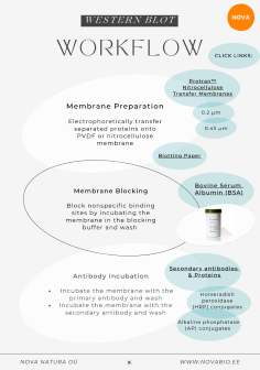
The Western blotting process involves several steps from membrane preparation to detection & visualization. We have prepared a guideline to help you navigate through this process.
Brochure:
Tips & Tricks:
Sample preparation:
Ensure proper sample handling and storage to maintain protein integrity.
Use fresh and high-quality samples to avoid degradation and loss of protein.
Consider protein extraction methods suitable for your sample type.
Optimize protein concentration for loading by using a protein assay, such as the Bradford or BCA assay.
Gel electrophoresis:
Choose the appropriate percentage of polyacrylamide gel based on the size of the target protein. Generally, lower percentage gels are suitable for separating larger proteins, while higher percentage gels are used for smaller proteins. Here is a general guideline for selecting the appropriate polyacrylamide gel percentage based on protein size: 4–40 kDa 20% 12–45 kDa 15% 10–70 kDa 12.5% 15–100 kDa 10% 25–200 kDa 8% NB!Acrylamide is a potent cumulative neurotoxin: wear gloves at all times.
Ensure proper sample loading and equal protein loading across lanes (Avoid overloading - A good practice is to load only 75% of the well volume to avoid spillover into adjacent lanes)
Include molecular weight markers to estimate the size of the target protein accurately.
Protein transfer:
Optimize transfer conditions (time, voltage, and buffer composition) based on the molecular weight of the target protein.
Use appropriate membranes (nitrocellulose or PVDF) based on the protein properties and detection method.
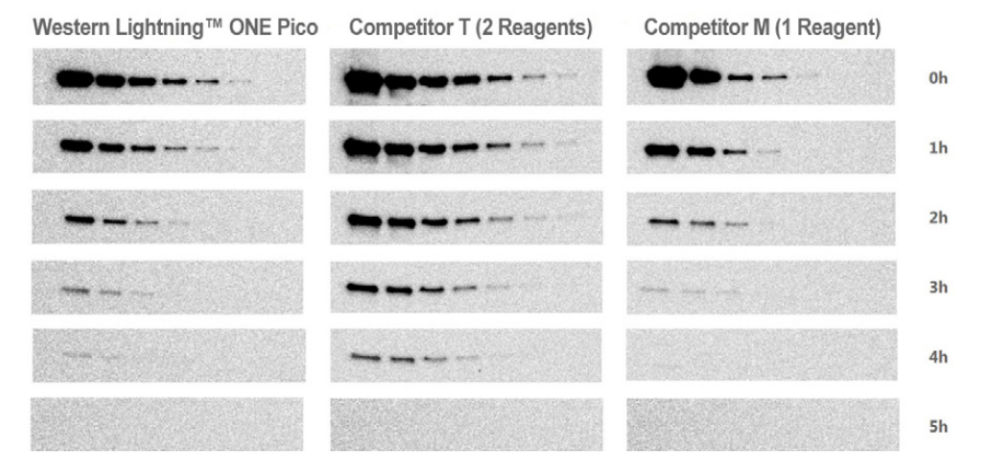
Explore different detection methods such as chemiluminescence or fluorescence-based detection.
To increase the signal intensity and save the antibody, see also: Western Lightning™ ONE – Premixed one-component chemiluminescent HRP substrate offering a range of protein sensitivity from mid picogram to low femtogram for more consistent results. Western Lightning™ – Two-component chemiluminescent HRP substrate for reliable and sensitive protein detection.
For the convenience of direct colorimetric visualization of results without the need for a film or imaging instrument, count on the DAB and BCIP/NBT substrates for use in chromogenic assays.
1) DAB to detect horseradish peroxidase (HRP)
2) BCIP/NBT to detect alkaline phosphatase (AP)
6. Data analysis:
Use appropriate quantification software to analyze band intensity, such as ImageJ or similar.
Normalize protein expression levels to a loading control (e.g., GAPDH or β-actin) for accurate comparisons.
Perform statistical analysis to determine significant differences between samples.
7. Troubleshooting:
If you encounter low signal or high background noise, optimize blocking conditions, antibody concentrations, and washing steps.
Verify the specificity of antibodies by performing positive and negative controls.
Check the integrity of the transfer by staining the membrane with Ponceau S before blocking.
Remember, western blot analysis can be a complex technique, and optimization is key to obtaining reliable and reproducible results. It may require several rounds of experimentation to achieve optimal conditions for your specific experiment.